Hyperpigmentation is rarely a single disorder. The visible mark (whether brown, grey, or violet) is the endpoint of a tightly regulated melanocyte response that runs through tyrosinase, melanosome transfer, and dermal pigment trapping. Effective treatment requires understanding which step in the cascade produces the specific lesion, because each step has its own active ingredients.
Melanocyte biology and tyrosinase
Melanocytes sit at the basal layer of the epidermis. Each cell extends dendrites to roughly thirty neighbouring keratinocytes, packaging melanin into membrane-bound organelles called melanosomes and transferring them through phagocytic uptake. The rate-limiting enzyme in melanogenesis is tyrosinase, which converts L-tyrosine to L-DOPA and then to dopaquinone, the chemical entry point of the entire pigment pathway.
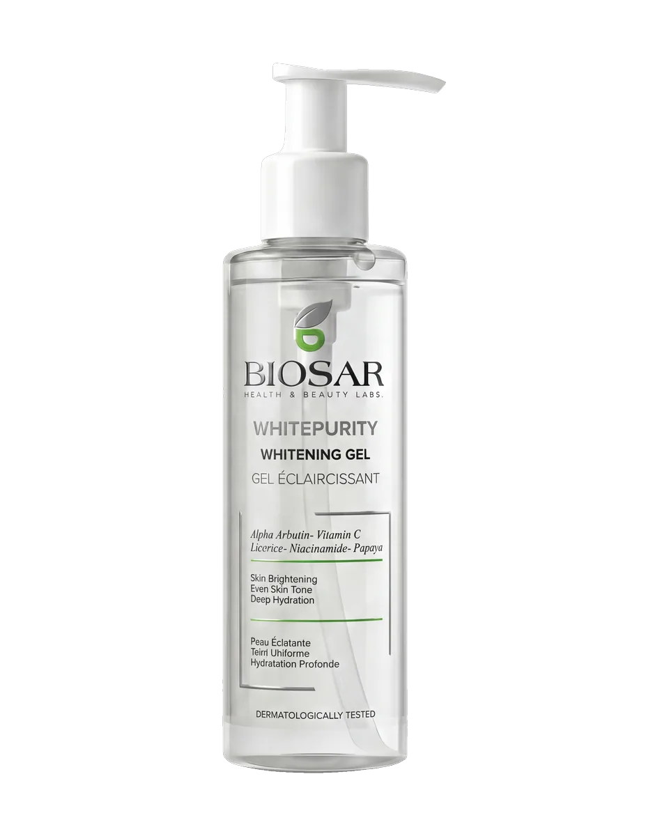
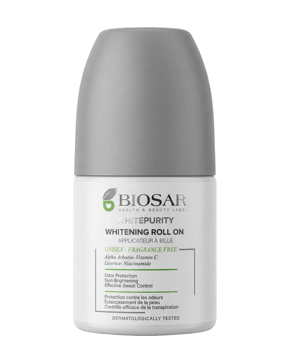
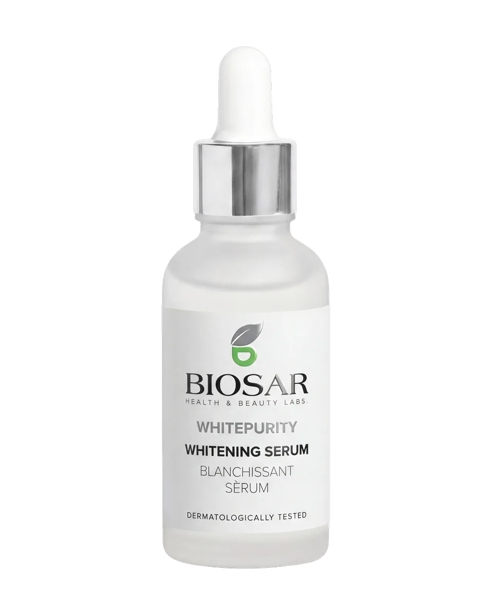
Almost every effective topical pigmentation active inhibits tyrosinase or interrupts melanosome transfer. Vitamin C reduces dopaquinone back to L-DOPA. Alpha-arbutin and kojic acid bind the active site of tyrosinase. Niacinamide does not touch tyrosinase but blocks the transfer of finished melanosomes from melanocyte to keratinocyte, a mechanism that makes it complementary to tyrosinase inhibitors rather than redundant. The clinical pattern is consistent: layered actives outperform any single agent in controlled trials.
Post-inflammatory hyperpigmentation
PIH is the pigment that follows inflammation: acne, eczema, ingrown hair, cosmetic procedures, or thermal injury. The inflammatory cytokine cascade (IL-1, prostaglandins, leukotrienes) directly activates melanocytes in the lesion bed. The result is a sharply demarcated brown or violet mark that can persist three to twelve months after the original insult resolves.
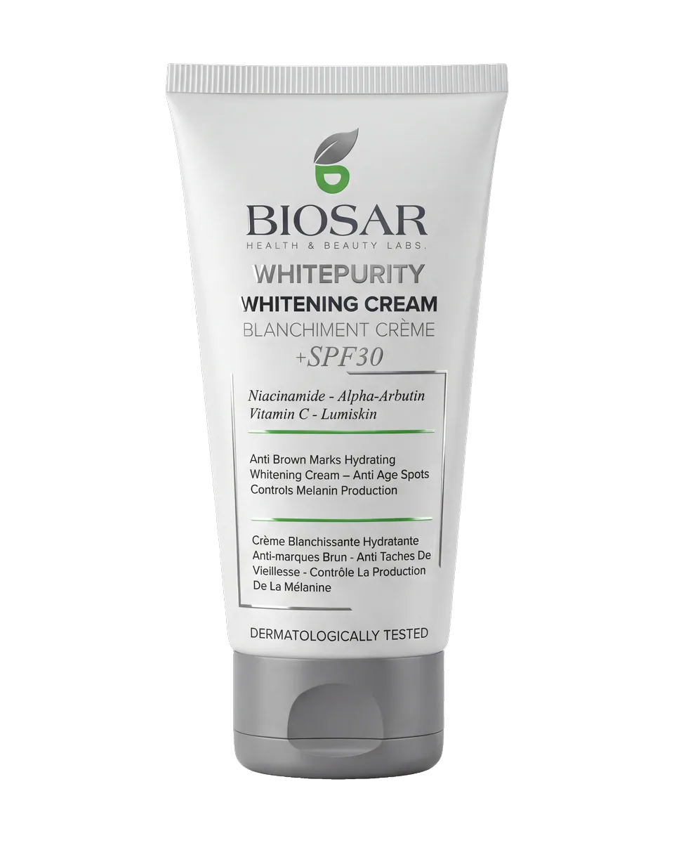
Two factors lengthen PIH duration in deeper skin tones. First, Fitzpatrick III–V melanocytes produce larger, more numerous melanosomes. The same inflammatory signal yields a deeper mark. Second, ongoing UV exposure renews tyrosinase activation each day, so the mark fades only as fast as fresh pigment production drops below the natural turnover rate. Daily SPF and a tyrosinase inhibitor in the morning, paired with a niacinamide-rich serum at night, is the standard published protocol.
Melasma and hormonal pigmentation
Melasma presents as symmetric brown or grey-brown patches across the cheeks, forehead, upper lip, and chin. It is strongly hormone-driven. Pregnancy, oral contraceptives, and hormone replacement each elevate oestrogen and progesterone, which upregulate melanocyte proliferation and tyrosinase activity. Genetic susceptibility runs strong in deeper skin tones.
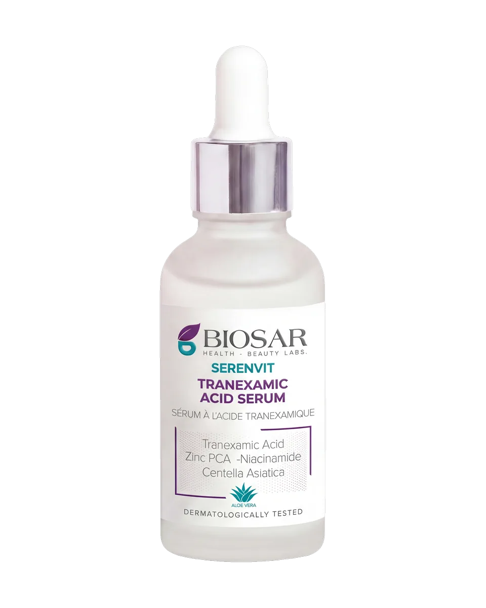
Melasma is more difficult to clear than PIH. The pigment often deposits not only in the epidermis but also in dermal macrophages, where topical actives reach poorly. Tranexamic acid (both topical and low-dose oral, where prescribed) is the most studied modern intervention, with multiple controlled trials showing significant fade over three months (Source: Taraz et al., Dermatol Ther 2017). Vitamin C, alpha-arbutin, and azelaic acid are first-line topical complements. Melasma always rebounds without rigorous daily SPF, including a tinted mineral filter that blocks visible light. Visible light alone provokes melasma in pigmented skin, even with strong UV protection.
Why SPF is non-negotiable
Across all three pigmentation types, UV exposure is the single largest accelerant. UVA renews tyrosinase activation daily. UVB triggers immediate pigment darkening. Visible light, particularly the blue-violet 400–450 nm band, drives melasma even when UV is fully blocked. Mineral filters with iron oxides cover this gap and remain the preferred photoprotection for pigmentation patients. A pigmentation routine without daily broad-spectrum SPF is a routine that will fail. The clinical evidence on this point is unambiguous and decades old.